Metastasis dependancy on the primary tumor?
Written by Ivo Mitsiev at in "Hepatobiliary Surgery" and also in "Cases", "Surgical Oncology".Recently I had an interesting case with a weird behavior of a suspected metastasis after resection of the primary tumor.
Description
A 43 years old gentleman came in July 2008 to the Medical Service with nausea, vomiting and abdominal pain. The workup showed a tumor in the 3rd portion of the duodenum as well as a big mass in the right liver lobe, so the IM guys asked us to take a look. They performed two attempts to biopsy the masses which showed no tumor. This was the reason for the decision to resect the tumor.
Here are the interesting parts of the initial imaging. They show the duodenal mass, as well as the big mass in the right liver lobe. The liver finding could be clearly identified in the PET scan also, without evidence for any other metastasis.


In July 2008 an exploratory laparotomy with a pylorus sparing modification of the Whipple procedure was performed, the right liver was not resected. The pathology result showed GIST.
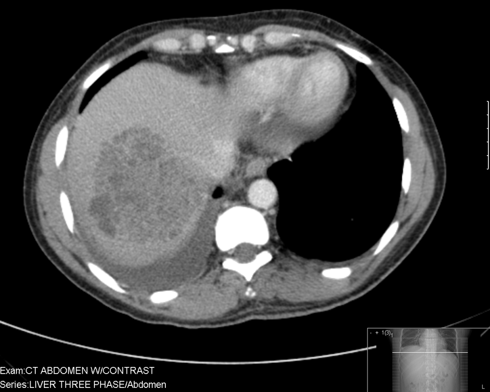
Some two weeks postoperatively the patient was admitted because of abdominal pain and nausea. A CT scan showed no bowel obstruction. An interesting finding was though the shrunk liver mass wich was now localized only to segment 8:

The patient could be discharged and admitted four weeks later for the planned right hemihepatectomy. Intraoperatively no significant liver tumor could be palpated. The right lobe was resected and segment 8 was opened where a small mass of scar and probably tumor could be seen.
The pathology reading came and... no tumor was found in the liver!
The patient received therapy with imatinib PO and was discharged in good shape.
Discussion
The good result suggesting a good prognosis raised though the obvious question what happened and what made the liver mass disappear some weeks after it was shown to actually be huge.
Possible scenarios are: liver abscess, liver metastasis of the GIST and a necrosis.
Even though the patient was initially admitted with some fever and leukocytosis, the following course makes the scenario with the liver abscess not that probable. The fever improved quick and spontaneous, the patient felt soon much better which would have been unusual findings in a liver abscess with this volume.
A liver metastasis of the GIST was the first idea everyone had after seeing the pictures. I admit, nothing was found on the biopsies. Though negative findings on biopsies give usually no information to a good surgeon.
Unfortunately metastases do not disappear after resection of the primary tumor. And I have no idea, how this could have been possible. The metastasis was countinuously fed and dying in starvation after resection of the primary tumor? Come on... this is not seriuos.
I think that the third scenario is the most probable. Tumor emboli spread in the liver could be a possible explanation for a liver necrosis which shows as a liver mass. This could explain also the abdominal pain which was one of the major symptoms for the initial presentation of the patient. The 6 weeks between the finding of the big liver mass and the liver resection with no tumor on histology are actually enough for the liver to regenerate. The CT scan performed in the middle of this time period shows the shrunk of this liver mass which would correlate more to the idea of the necrosis than of the abscess.
I wish, we could get this result in metastasis finding after resection of primary tumors.