Brenner Tumor
Written by Ivo Mitsiev at in "General Surgery".In 1907 Fritz Brenner published his thesis "Das Oophoroma folliculare".
F. Brenner: Das Oophoroma folliculare. Frankfurter Zeitschrift für Pathologie, München, 1907, 1: 150-171)
He described what was later called "Brenner Tumor".
These tumors are rare epithelial ovarian tumors that account for 1-2% of all ovarian neoplasms. They are divided into benign, borderline or proliferative, and malignant neoplasms. In the vast majority of cases, these lesions are benign. Tumors of borderline malignancy are less frequent and only about 1% of Brenner tumors are malignant.
The diagnosis is usually made incidentally on physical examination of pelvis or lower abdomen as well as on abdominal surgery (for other reasons). The tumors can grow without any symptoms and diameters of up to 30 cm were described. They often coincide with a mucinous cystadenoma or dermoid cyst and are most common in the fifth to sixth decades of life.
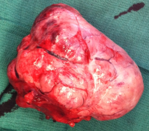
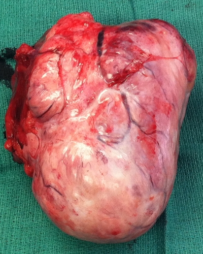
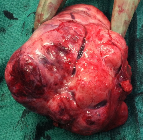
Today I found one in a 67 years old brain-dead lady who was intended to undergo a multi-organ donor surgery. The tumor had its origin from the left ovary pushing the sigmoid colon to the right side having embedded the left ureter. It was adherent to the uterus and its diameter was about 15 cm.
In addition a tumor was found in liver segment 3 which was completely excised and sent to pathology for frozen section. Grossly it looked very malignant but it was reported to be just focal nodular hyperplasia (FNA). The pathologist could not exclude malignancy of the Brenner tumor. Here are some pictures of it:


A quick review of the literature in PubMed did not show more than a lot of all kind of case reports, mostly about malignant Brenner tumors as they are quite rare. I just decided to summarize the facts I found while refreshing my memory about those kind of tumors.